




Bioimaging data analysis: from zero to machine learning hero

Bioimaging data analysis: from zero to machine learning hero
19 October 2026, 9h00 - 17h00
20 October 2026, 9h00 - 17h00
21 October 2026, 9h00 - 17h00
General context
Bioimaging Data Analysis: From Zero to Machine Learning Hero is a three‑day, project‑style course that guides participants through the full journey of understanding, exploring, and analyzing bioimaging data. Beginning with common pitfalls in image acquisition and an accessible introduction to machine learning, participants progressively build practical skills using powerful open‑source tools. Hands‑on sessions with Napari, QuPath, ilastik, and Cellpose provide an interactive foundation for image visualization, segmentation, and quantitative analysis, while lunch‑and‑learn moments introduce broader concepts such as emerging machine‑learning approaches and the Bioimaging Model Zoo.
In the final stretch, participants dive into state‑of‑the‑art deep‑learning tools, including SAM, Empanada, and Napari‑based neural network workflows, before shifting perspective toward responsible and reproducible science. Day three focuses on image ethics, FAIR data principles, and personalized project work in a “Bring Your Own Data” session—allowing each participant to apply new skills directly to their own research questions and leave the course with practical, actionable experience.
This is an event organized as 'RACE event', work package 3 of the RACE project funded by the European Union under Horizon Europe (Project no 101059801).
Read more about RACE at https://www.iimcb.gov.pl/en/race/
Funded by the European Union. Views and opinions expressed are, however, those of the author(s) only and do not necessarily reflect those of the
European Union or the European Research Executive Agency. Neither the European Union nor the granting authority can be held responsible for them.
Explain common pitfalls in bioimage acquisition and evaluate how they affect downstream analysis quality.
Operate core functionalities of Napari, QuPath, ilastik, and Cellpose to visualize, segment, and quantify biological images in a reproducible manner.
Compare classical image‑analysis approaches with machine‑learning–based workflows, selecting appropriate methods for different bioimaging tasks.
Apply pixel‑ and object‑classification workflows in QuPath and interpret the resulting segmentation outputs for biological relevance.
Execute deep‑learning‑based segmentation tools such as SAM and Empanada, and assess their performance using appropriate evaluation criteria.
Discuss best practices in image ethics and apply FAIR data principles when preparing imaging data for sharing, publication, or reuse.
Design and implement a basic analysis pipeline for their own dataset (“Bring Your Own Data”), demonstrating problem‑solving, tool selection, and critical interpretation of results.
Fundamental understanding of microscopy data.
Students should know the essentials of how biological images are acquired (e.g., channels, resolution, noise, file formats) to make sense of topics such as acquisition pitfalls, segmentation workflows, and model performance.
Trainers
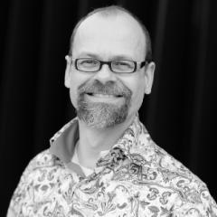
Sebastian Munck
Laura Murphy
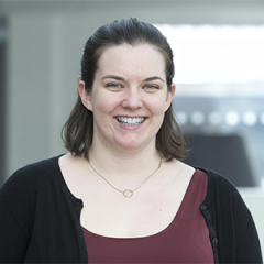
Laura Murphy completed her PhD in Neurobiology at the University of Edinburgh in 2017. Following her doctoral studies, she transitioned to her current role as a Bioimage Analyst for the Institute of Genetics and Cancer Advanced Imaging Resource. In this capacity, Laura supports researchers by developing strategies to extract precise quantitative information from complex microscopy data, bridging the gap between raw imaging and biological insight.
Benjamin Pavie

Tomasz Wegierski
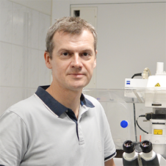
Since 2021, Tomasz Węgierski has served as the Head of the Microscopy Facility at the IN-MOL-CELL infrastructure, located at the International Institute of Molecular and Cell Biology (IIMCB) in Warsaw, Poland. The facility provides services and access to advanced light and electron microscopes, as well as flow cytometers, for eleven laboratories at IIMCB and for external users from the University of Warsaw and several research institutes.
From 2009 to 2021, he worked at IIMCB as a senior scientist and facility staff member. Prior to that, he completed his postdoctoral training in Freiburg, Germany, from 2003 to 2009, following the completion of his PhD in 2002 at the University of Basel, Switzerland. His doctoral research focused on proteins involved in pre-rRNA processing in the yeast Saccharomyces cerevisiae.
Tomasz Węgierski specializes in light microscopy imaging techniques, image processing and analysis, and has a strong interest in optical instruments, ranging from microscopes to telescopes.
Ann Wheeler
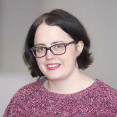
Dr. Ann Wheeler, is a leading figure in the field of advanced microscopy. Dr. Wheeler heads the Advanced Light Microscopy Facility at the Institute of Genetics and Cancer, University of Edinburgh. Since 2014, her leadership has transformed the facility into a hub of innovation, equipped with cutting-edge microscopes, including those for super-resolution techniques like SIM and STORM, integral to the Edinburgh Super Resolution Imaging Consortium (ESRIC) and Bioimage analysis.
A Fellow of the Royal Microscopical Society, Dr. Wheeler also contributes significantly to initiatives such as the European Light Microscopy Initiative and EuroBioImaging. She is the driving force behind Edinburgh Bioimaging and leads the University of Edinburgh Bioimaging UK Nodelet.
Dr. Wheeler's academic path began with a MBioch in Molecular Biochemistry from the University of Oxford, followed by a PhD at the University of London, conducted at the prestigious Ludwig Institute for Cancer Research. Her professional journey includes managing microscopy facilities at Queen Mary, University of London, and engaging in impactful postdoctoral research at Imperial College London and Scripps Research Institute.
With a focus on advancing imaging research, super-resolution techniques, and cytoskeletal studies, Dr. Wheeler's work has significantly contributed to our understanding of cellular dynamics and cancer research. Her extensive publications underscore her commitment to pushing the boundaries of what microscopy can achieve.
Program
Welcome and short introduction of instructors and participants
Pitfall of the acquisition
What has machine learning ever done for us?
Lunch (provided)
Napari Introduction
Coffee break (provided)
Napari Part II
Napari Part III
Coffee break (provided)
QuPath - pixel classifier/ object classifiers (a fast intro)
Lunch (provided)
Ilastik - towards machine learning
Coffee break (provided)
FAIR Imaging resources and image ethics (publications)
Deep learning with Napari : Noise2Void/Micro-SAM/NNInteractive/Empanada
Coffee break (provided)
Bring your own data (present your challenges)
Lunch (provided)
Bring your own data (working on the examples with experts)
Practical info


19 October 2026 - 21 October 2026
IIMCB - International Institute of Molecular and Cell Biology in Warsaw
Hankiewicza 2
02-103 Warsaw
Poland
19 October 2026 - 21 October 2026
IIMCB - International Institute of Molecular and Cell Biology in Warsaw
No public transport information has been provided for this location.
19 October 2026 - 21 October 2026
IIMCB - International Institute of Molecular and Cell Biology in Warsaw
19 October 2026 - 21 October 2026
IIMCB - International Institute of Molecular and Cell Biology in Warsaw
No parking information has been provided for this location.
19 October 2026 - 21 October 2026
IIMCB - International Institute of Molecular and Cell Biology in Warsaw
No contact information for this location has been provided.