




Intravital Microscopy Training

Intravital Microscopy Training
15 November 2024
General context
This one-day training day in Intravital Imaging techniques and setups will aim to showcase a variety of intravital imaging techniques and is preceded by the 4th Day of Intravital Microscopy. The day will start out with a small seminar series in the morning explaining and breaking down intravital imaging surgical techniques as well as imaging setups used. The training day will then conclude in the afternoon with live demonstrations of abdominal, thoracic and cranial imaging windows and imaging on our 2 inhouse 2-photon Leica SP8 (upright) and DIVE (inverted) microscopes as well as a novel intravital aiming system IVM-CMS3 demoed by IVIM Technologies.
This course is intended for researchers who want to learn more about possible intravital imaging techniques and take a deep dive into the set-ups available at the VIB Center for Cancer Biology Intravital Imaging Expertise Center.
Trainers
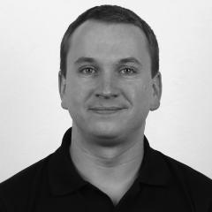
Max Nobis
https://ccb.sites.vib.be/en/expertise/intravital-imaging
Max Nobis heads the Intravital Imaging Expertise Center at the VIB-KU Leuven Center for Cancer Biology in Leuven, Belgium using a wide array of intravital imaging techniques to address a broad range of research questions in the fields of immunology, metabolism, development, cancer metastasis and neurobiology. He has 12 years of experience in intravital imaging techniques and advanced 2-photon microscopy (46 publications, h-index 26). He performed his PhD research the CRUK Beatson Institute, Glasgow, UK establishing the use of FRET biosensor mice to dissect cancer progression and spatiotemporal treatment mapping using optical imaging windows (Nobis et al. 2013 Cancer Research). During his post-doctoral studies at the Garvan Institute of Medical Research in Sydney Australia, his work has further centered around the use and characterization of novel FLIM-FRET biosensor mice in metabolism, infection, inflammation, cancer and development research (Nobis et al. 2017 Cell Reports, Floerchinger et al. 2021 Cell Reports, Conway et al. 2023 Science Advances).
Michael Gerlach
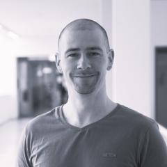
Michael Gerlach is an expert intravital imaging scientist having received his Diploma at the TU Dresden and completed his PhD at the Center for Regenerative Therapies in Dresden, where he explored longitudinal imaging of pancreatic islets in the AEC. He went on to do his post-doc at the Department of Nephrology at the university hospital in Dresden and works as of 2018 as an application specialist for intravital imaging at the Core Facility Cellular Imaging located at the Medical Faculty “Carl Gustav Carus” in Dresden.
Michael Kuligowski
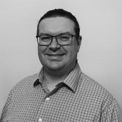
Michael Kuligowski is a scientist who specializes in Intravital imaging to try and understand dynamic biological processes that occur during pathology of disease. Michael did a Bachelor of Science at The University of Melbourne, before completing his PhD at Monash University investigating leukocyte and platelet recruitment to the glomerular capillary beds within the kidney. He then did a post doc in Boston, at Harvard Medical School where he investigated influenza trafficking in lymph nodes, visualizing the virus enter, bind to, and get passed from macrophages to dendritic cells during the immunization process. Michael then returned to Australia working at The Centenary Institute visualizing immune mediated organ and graft region in skin grafts and islet cells.
A few years ago, Michael joined the microscopy core facility at The University of Sydney, where he manages a microscopy facility, but specializes in assisting researchers set up and then perform intravital imaging experiments.
Asli Ayaz
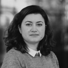
Asli Ayaz has been a senior scientist at VIB and Neuro-Elecetronics Research Flanders (NERF) since 2019. In Jan 2024, she took on the expert neuroimaging scientist role within the In Vivo Neurophysiology Expertise Unit. She obtained her PhD degree in Neurobiology from the University of California Irvine. Her past research experience extends to both the visual and somatosensory systems and computational approaches. As a postdoctoral researcher at the University College London (UCL) and the University of Zurich, she demonstrated principles of sensorimotor integration in visual cortex neurons (Ayaz et al., 2013, Current Biol, Saleem et al., 2013, Nat Neuro) and across layers of somatosensory cortex (Ayaz et al., 2019, Nat Comm), respectively. Most recently, she was awarded a Marie Curie Individual Fellowship (2021) to study large-scale circuit mechanisms of multisensory information selection. She has extensive experience in state-of-the-art in vivo imaging and neural recording techniques, behavioral approaches, and computational skills that are vital for the in-vivo characterization of network dynamics. She aspires to disseminate in-vivo neural circuit manipulation and neural recording approaches to the non-expert scientific community and facilitate deciphering circuit function anomalies in in vivo models of brain disorders.
Ben Vermaercke
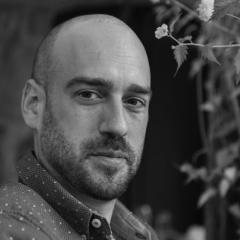
Lara Roosen
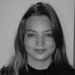
Jef Evenepoel
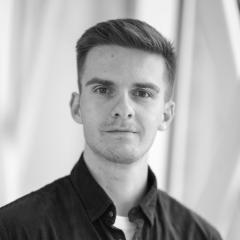
Intravital Imaging Expertise Center & Laboratory of Intravital Microscopy and Dynamics of Tumor Progression
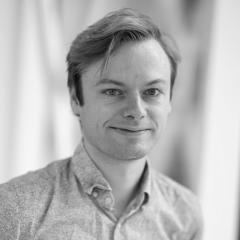
Lander de Herdt
Program
Morning theoretical session
Introduction
Max Nobis, Intravital Imaging Expertise Center VIB-KU Leuven Center for Cancer Biology
Intravital Imaging of organs in the peritoneum and thorax
Max Nobis, Intravital Imaging Expertise Center VIB-KU KU Leuven Center for Cancer Biology
Abdominal windows for longitudinal imaging of kidney (patho)- physiology
Michael Gerlach, Core Facility Cellular Imaging, Medical Faculty Carl Gustav Carus, Dresden, Germany
Coffee break
Lymph Node imaging in mice, problems and solutions
Michael Kuligowski, Sydney Microscopy & Microanalysis, Brain and Mind Center, University of Sydney, Australia
In vivo functional brain imaging approaches and applications in animal models
Asli Ayaz, In Vivo Neurophysiology Expertise Unit, NERF, VIB-KU Leuven
Optimized cranial window implantation for subcellular and functional in vivo imaging
Ben Vermaercke, VIB-KU Leuven Center for Brain & Disease Research
Lunch
Afternoon practical session
Groups of max. 7 people will rotate through the following stations
Lara Roosen – Cranial Window Set up
Jef Evenepoel – Abdominal Window Set up
Lander de Herdt – Lung Window Set up
Practical info


15 November 2024
Leuven - Campus Gasthuisberg
Herestraat 49
3000 Leuven
Belgium
15 November 2024
Leuven - Campus Gasthuisberg
Leuven train station is approximately 4 km from the campus Gasthuisberg. Buses depart very regularly to the Gasthuisberg Campus from Leuven train station.
For tickets and up to date schedules visit website De Lijn or use the De Lijn App.
Exit at the very last bus stop ‘Gasthuisberg Campus) make sure not to exit at ‘Gasthuisberg Kliniek =
hospital)
Once you get off the bus, go to your right and go up the street until you see stairs to your left. Climb the stairs all the way up between two buildings. Look for the signs containing blue circles and white numbers and follow them all the way from 2 up to number 12. You will cross several buildings and the walk will take max. 10 minutes. When you reach number 12, you have reached O&N 5, where you can find the reception.
15 November 2024
Leuven - Campus Gasthuisberg
The Gasthuisberg campus is west of the Leuven ring. Our buildings are situated behind the hospital (see black arrow near ‘RING NOORD’ on the map). If you come from the city center, you will arrive on campus from the east side (taking Mgr. Van Waeyenberglaan and roundabout Het Teken). If you come from Heverlee or Campus Arenberg, you will arrive from the west side (taking the Celestijnenlaan and Taborweg). When you arrive uphill at the intersection with the Rega Institute and the Red Cross building, you need to take the road down to the bicycle parking at Rega Institute (see map) where you can park. Next, follow the ‘Onderwijs & Navorsing blue route’ description below (start point is #9).
15 November 2024
Leuven - Campus Gasthuisberg
You arrive via the E314 highway – EXIT 16
After 200 m, keep left to drive underneath the roundabout and into the tunnel.
Keep left and follow the arrows to Parking West.
You arrive via the Leuven ring road
At the roundabout, take the exit towards ‘Spoed / Ziekenhuis’.
Follow the ring around Campus Gasthuisberg and pass the entrance of SPOED.
Continue right until you arrive at a second roundabout.
Follow the directions for E314.
Near the Fire Brigade Station, make a U-turn and follow the directions for ‘Ziekenhuis’.
Keep left and drive downhill, underneath the roundabout and into the tunnel.
Keep left and follow the arrows to Parking West.
Next, leave the parking at level +1 using the big stairs near the vending machines.
Follow the red carpet and arrows to 'Onco Dagcentrum' to get to the O&N buildings. Take the stairs on your left after exiting Parking West. You are now in front of the O&N4 building; on the left you will find O&N5.
15 November 2024
Leuven - Campus Gasthuisberg
ON5 - auditorium